
Ventricular pressure-volume (PV) relationships are a well established and comprehensive means for assessing cardiac function across a range of animal models.
The technique was originally developed for use in large mammals and humans; however, through the miniaturization of conductance catheters and optimization of surgical procedures, ventricular PV analysis has successfully been translated to small rodents, opening up the opportunity to assess cardiac function in genetically engineered mouse models and drug treatment studies.1
Correct placement of the pressure-volume catheter in the ventricle is essential for collecting high-quality, accurate data. There are two main surgical approaches for inserting a pressure-volume catheter into the left ventricle (LV); the carotid (closed-chest) and apical (open-chest) approach.

Below, cardiovascular physiologist DeWayne Townsend (DVM, Ph.D.) from the University of Minnesota Medical school, discusses the advantages and limitations of the carotid and apical approach and the appropriate situations to use them.
Related: Tips and tricks for measuring PV loops in mice
Carotid approach (closed-chest)
The carotid approach is a closed chest approach, which involves inserting the PV catheter through the right carotid artery into the LV. Below we will outline some key advantages and limitations to consider when using this surgical approach.
Advantages
- Closed chest approach - The main advantage of the closed-chest carotid approach is that it prevents major trauma from a thoracotomy, which can influence cardiac physiology. As such, the carotid approach is more suited to prolonged experiments like drug testing, that require the mouse to be catheterized for a longer period of time. 1,2
- Arterial pressure recordings are easily obtained - Because the catheter is advanced through the right carotid artery, arterial pressures can easily be obtained at the beginning and end of the experiment. This is particularly useful when studying disease models like heart failure, where the total peripheral resistance (TPR) is an important measure. 1
- Ventilation is not required - Because the chest is closed, ventilation is not required. However, it’s important to note that ventilation allows for greater respiratory control and aids in the analysis of your PV data. Therefore, some people may still consider ventilating their animals when using the carotid approach.
Limitations
- Catheter placement is defined by aortic anatomy - Because the catheter is directed down through the aorta into the LV, once inserted, adjusting the position can be difficult. This can pose problems if your catheter placement isn't quite right or there is an internal cardiac structure occluding or constricting down on the catheter.
- Laparotomy required to occlude vena cava - To perform an inferior vena cava (IVC) occlusion using the carotid approach, a second surgical incision must be made in the abdomen to expose the vena cava.
- Potential for outflow track obstruction in smaller hearts - Another caveat of the carotid approach is the potential for outflow track obstruction. When working with smaller mice hearts, the size of the catheter relative to that of the aorta can impact the outflow of blood from the ventricle, impacting on the hemodynamics of the heart by causing an increase in afterload. 3
- Not appropriate for disease models of extreme hypertension or severe atherosclerosis.
See Pál Pacher et al. (2008) Nature Protocol for more information.
Related: See Millar's full range of PV Conductance Catheters for small and large animal models.
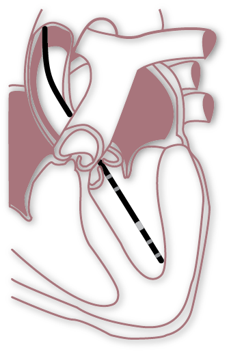
Apical approach (open-chest)
The Apical approach (or ventricular apical stab method) is a open chest approach. For this, a small incision is made through the diaphragm of the animal to expose the apex of the heart. A 25 gauge needle is then used to create a stab incision at the center of the apex, through which the catheter will be inserted.2,3 The apical approach is considered the least challenging of the two techniques, as proper placement of the catheter in the ventricle is more easily achieved.
Advantages
-
Catheter placement can be optimized - One of the main advantages of the open-chest apical approach is it allows for greater control over the catheter position and orientation within the ventricle.
- No outflow track obstruction - As the catheter is inserted through the base of the ventricle, you don't need to worry about outflow track obstructions.
- No additional surgery is required for IVCO - As mentioned, the apical approach is an open-chest technique, so access to the vena cava for modulating cardiac loading is easily achieved. Find out more: Tips for modulating cardiac loading through IVC occlusions and abdominal compressions.
Limitations
- Requires ventilation - Because you’re opening up the chest of the animal, ventilation is required. However, ventilation isn’t always considered a disadvantage. It can give you greater control over the health of the animal and aid in data quality. This is why some researchers chose to ventilate their animals even when using the carotid approach.
Find out more - Dealing with respiratory artifacts from positive pressure ventilation »
- Extensive surgical manipulation - The apical approach requires significant modification of the chest wall in order to access the heart for catheterization, which can influence the hemodynamic properties of the cardiovascular system.

- Potential to damage the myocardium - One major challenge when starting out with the apical approach is avoiding damage to the heart when opening the chest cavity. Over time, as you become more proficient in the technique, this will become less of an issue.
Hopefully, this summary has helped you decide which is the best surgical approach for your PV research. For more information about PV loop data acquisition and analysis, check out Dr. DeWayne Townsend and Dr. Adam Goodwills' full webinar on tips and tricks for cardiac PV loop analysis - or get in contact with your nearest support specialist - we'd be happy to help!
Read More:
Troubleshooting your PV catheter - three common problems and how to fix them
Understanding ventricular pressure-volume catheter calibrations and experimental design
Optimizing your pressure-volume recordings in LabChart
LabChart – PV Loop analysis for left and right ventricle studies
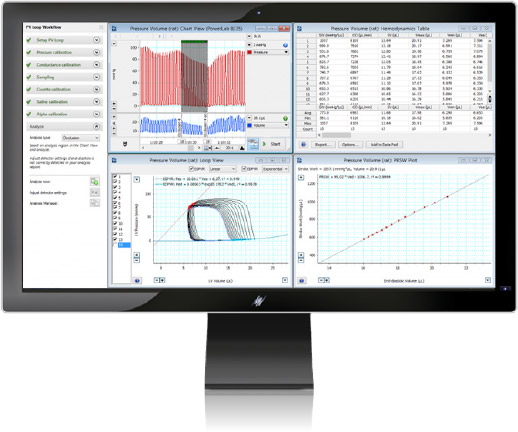
LabChart's PV Loop Analysis Module is specifically designed for the analysis of in vivo ventricular pressure-volume data in small and large animals, or ex vivo, using working heart systems.
The PV Loop Analysis Software offers smart presets for different animals and streamlined workflows, guiding you step-by-step from calibration through to analysis.
Find out more »
References:
1. Pacher, P., Nagayama, T., Mukhopadhyay, P., Bátkai, S., & Kass, D. A. (2008). Measurement of cardiac function using pressure-volume conductance catheter technique in mice and rats. Nature protocols, 3(9), 1422–1434. https://doi.org/10.1038/nprot.2008.138
2. Abraham, D., & Mao, L. (2015). Cardiac Pressure-Volume Loop Analysis Using Conductance Catheters in Mice. Journal of visualized experiments : JoVE, (103), 52942. https://doi.org/10.3791/52942
3. Townsend D. (2016). Measuring Pressure Volume Loops in the Mouse. Journal of visualized experiments : JoVE, (111), 53810. https://doi.org/10.3791/53810